Anatomical models
MIT engineers are hoping to help doctors tailor treatments to patients’ specific heart form and function, with a custom robotic heart.
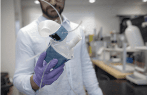
[Image courtesy of Melanie Gonick, MIT]
3D printing is allowing device developers and surgeons to build personalized models of organs, bones and other bodily structures. These can be used for training, education, pre-operative planning or to test new and improved device prototypes.
MIT engineers are 3D printing individualized heart replicas to test how a particular valve implant will affect the performance of a specific patient’s heart.
“We’re not only printing the heart’s anatomy, but also replicating its mechanics and physiology,” MIT mechanical engineering professor Ellen Roche said. “That’s the part that we get excited about.”
Meanwhile, Stratasys and Ricoh are partnering on 3D-printed, personalized anatomic models for clinical settings. The on-demand models provide specific visualizations of a patient’s anatomy to allow practitioners to plan and practice complex surgeries and improve communication between medical staff, the patient and families.
Next>>