Hologic designed its Genius cytology technology for more efficient and accurate review of cervix cell samples — and there’s more to come.

Hologic says its Genius cytology technology reduces false negatives of high-grade squamous intraepithelial and more severe lesions by 28% compared to microscopic review. [Photo courtesy of Hologic]
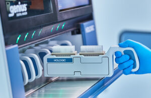
Hologic‘s Genius cytology system uses new scanning technology and artificial intelligence to flag cervical cancer cells and pre-cancerous lesions.
Hologic won a de novo classification in January 2024 for its Genius Digital Diagnostics System and Genius Cervical AI algorithm for cervical cytology. Besides replacing Hologic’s ThinPrep Imaging System — used for the majority of cervical cancer screenings in the U.S. — the technology behind the Genius system could one day also help screen for other kinds of cancer like bladder cancer as well as infectious organisms, Hologic VP of R&D/Innovation Mike Quick said in an interview.
“It really has the potential of being applied to a variety of other disease states,” Quick said. “We’re excited about building out that roadmap and what that future state can look like. It really is more about a platform than it is as a single instrument that’s a replacement just for cervical cancer screening. … Candida, trichomonas, herpes can all be identified on the Genius platform using the same AI-type algorithms.”
Taking pictures of microscopic slides for analysis is not a new idea. Quick — who trained and worked in cytology before joining Hologic in 2007 — said he has copies of some publications looking at image analysis for cytology dating back to the 1950s. In the past 15 years or so, however, there’s been greater adoption of the concept in pathology than cytology.
“When we first started looking at this at the start of this project in about 2015, what we found is that there were real technological limitations of why that space hadn’t evolved,” he said.
The fundamental difference is that pathology images use flat tissue sectioned with a razor blade on a microscope slide for very thin uniformity across the entire material. But with cytology, they’re whole cells in three dimensions on the microscope slide.
“You’re taking a much deeper thickness of the material, almost like the difference between a chest X-ray and a CT scan,” he said.
Developing the Genius Digital Diagnostics System scanner

Hologic’s Genius Digital Diagnostics System scanner [Photo courtesy of Hologic]
Quick initially hoped that Hologic could develop the AI image analysis component and use existing scanners, but his team found those scanners weren’t suited for three-dimensional cells.
“Either they couldn’t get images in focus because of the multiple plane issue, or if they could, they had to do it by essentially taking multiple pictures: 13 to 15 pictures at a time of a field, and then move down through the material, taking pictures one at a time,” Quick said. “What was reportedly the best-in-class scanner on the market took about an hour and five minutes to scan one slide using that approach, which we knew would never be a commercially viable option.”
So Hologic developed what it calls volumetric imaging to simultaneously capture multiple images for a 3D image before converting it into 2D. It’s a similar approach to what’s called focus stacking in photography, where a photographer takes multiple shots of the same scene at different focal planes to combine them for crisp focus near and far.
“We’re able to take multiple planes at a single time and then reconstruct that in three dimensions, and then we we take that three-dimensional image and collapse it into a single two-dimensional image where each pixel that’s in focus in different planes is able to be combined into a flat image, with everything in focus. That’s really foundational. … It doesn’t matter how good your image analysis or any downstream analysis that happens is if your raw material isn’t the best that it can be,” he said.
Specimen slides being loaded into Hologic’s Genius Digital Diagnostics System scanner [Photo courtesy of Hologic]
Hologic aimed to scan each sample within two to three minutes, which led to another challenge: data transfer. Each scan generates about 100 GB worth of data, Quick said.
“Moving that amount of data that quickly gets into GPU optimization and efficient use of resources, doing things in parallel with scalability,” he said.
Developing the Genius Cervical AI algorithm

Cervical cells scanned and reviewed with Hologic’s Genius system [Photo courtesy of Hologic]
On the analysis side, Hologic developed an algorithm that uses machine learning to classify cells versus non cells, as well as deep learning to classify different kinds of cells. The algorithm is based on tens of thousands of images, Quick said, to provide more efficient and accurate review of the patient sample.
“It’s the approach of analyzing 70,000 or 80,000 cells on a single patient sample and distilling that down to 30 tiles, individual cells or cell groups that are representative of the entire cell sample,” he said.
Hologic says the technology reduces false negatives of high-grade squamous intraepithelial and more severe lesions by 28% compared to microscopic review.
“We don’t want to miss anything, but the high grades are the lesions that are one step away from becoming cancer,” Quick said.
Advice for other device developers
As a new technology, Hologic’s Genius system faced some common regulatory hurdles.
“Volumetric imaging — it’s new physics. It didn’t fit squarely into the boxes that the FDA had laid out in their guidance documents,” Quick said. “So how do you translate something that’s never been done before into the framework that the regulators are looking at?”
The Hologic team translated the technology for FDA reviewers through several informational meetings and then several pre-submission meetings.
“So much of that is about education,” Quick said. “The more that you can do on the front end — as laborious and time consuming as it can be — it certainly needs to happen so that you get alignment when you’re aiming for a bit of a moving target, particularly in this space.”

Hologic VP of R&D/Innovation Mike Quick [Photo courtesy of Hologic]
Quick offered some final advice for other device developers and manufacturers.
“You’ve got to have the tenacity and belief to see it through to the end,” he said. ” This was something we started pitching internally. It was the classic innovators dilemma of how do you invent and invest in something new when you’re the market leader in what you have today and need to vigorously defend what that is.”
“Building our internal advocates was as important as everything that we did externally to be able to move this into a project that our company invested in for the future,” he continued. “It’s been a long haul, but we definitely see the excitement and the enthusiasm, and literally have labs knocking down our door to talk about implementing it.”