
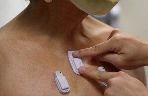
This soft, wireless device prototype can continuously monitor the body sounds inside and outside of a patient, including premature babies. [Photo courtesy of Northwestern University]
Northwestern University researchers have developed wearable devices for continuously monitoring the sounds made by a patient’s body, such as breathing, heartbeats and digestion.
The soft devices stick to a patient’s skin and use two high-performance, digital microphones to listen to sounds inside and outside the body. One of the microphones faces inside the patient, while the other faces outward and an algorithm separates external and internal sounds.
“Lungs don’t produce enough sound for a normal person to hear,” Northwestern Medicine thoracic surgeon Dr. Ankit Bharat said in a post at the university’s website. “They just aren’t loud enough, and hospitals can be noisy places. When there are people talking nearby or machines beeping, it can be incredibly difficult. An important aspect of our technology is that it can correct for those ambient sounds.”
Bharat led the team’s clinical research in 55 adult subjects, including 20 with chronic lung diseases. The researchers also tested their devices on 15 premature babies with respiratory and intestinal motility disorders.
“Lungs can make all sorts of sounds, including crackling, wheezing, rippling and howling,” Bharat said. “It’s a fascinating microenvironment. By continuously monitoring these sounds in real-time, we can determine if lung health is getting better or worse and evaluate how well a patient is responding to a particular medication or treatment. Then we can personalize treatments to individual patients.”
The researchers said their devices delivered clinical-grade accuracy and entirely new functions.
“Currently, there are no existing methods for continuously monitoring and spatially mapping body sounds at home or in hospital settings,” said Northwestern professor John Rogers, who led the device’s development. “Physicians have to put a conventional, or a digital, stethoscope on different parts of the chest and back to listen to the lungs in a point-by-point fashion. In close collaborations with our clinical teams, we set out to develop a new strategy for monitoring patients in real-time on a continuous basis and without encumbrances associated with rigid, wired, bulky technology.”
The team developed the devices “to provide highly accurate, continuous evaluation of patient health and then make clinical decisions in the clinics or when patients are admitted to the hospital or attached to ventilators,” Bharat said.
“A key advantage of this device is to be able to simultaneously listen and compare different regions of the lungs,” he said. “Simply put, it’s like up to 13 highly trained doctors listening to different regions of the lungs simultaneously with their stethoscopes, and their minds are synced to create a continuous and a dynamic assessment of the lung health that is translated into a movie on a real-life computer screen.”
The body sounds monitor device design
This wireless, wearable device has microphones that monitor body sounds inside of a patient. [Photo courtesy of Northwestern University]
The devices are 40 mm long, 20 mm wide and 8 mm thick. They’re encapsulated in soft silicone and contain a flash memory drive, battery, Bluetooth components, accelerometers and miniaturized microphones.
These components work together to map the flow of air in, through and out of the lungs; how cardiac rhythm changes when a patient is active or at rest; and how food, gas and fluids move through the intestines.
The devices are placed on babies just below the suprasternal notch at the base of their throats, or on the right and left chest.
“When placed on the suprasternal notch, the enhanced ability to detect and classify apneas could lead to more targeted and personalized care, improved outcomes and reduced length of hospitalization and costs,” paper co-author Dr. Wissam Shalish, a neonatologist at the Montreal Children’s Hospital. “When placed on the right and left chest of critically ill babies, the real-time feedback transmitted whenever the air entry is diminished on one side relative to the other could promptly alert clinicians of a possible pathology necessitating immediate intervention.”
When placed on a baby’s abdomen, the devices can listen for digestion issues, intestinal dysmotility and potential obstructions.
The devices also monitor and record noise levels around babies, which “offers immediate opportunities to address any sources of stressful or potentially compromising auditory stimuli,” Shalish said.
These noninvasive, continuous monitors can also collect data without disturbing the babies, many of which are smaller than a stethoscope and already technically challenging to monitor, said co-author Dr. Debra Weese-Mayer, chief of autonomic medicine at Ann & Robert H. Lurie Children’s Hospital of Chicago.
” These acoustic wearables provide the opportunity to safely and non-obtrusively determine each infant’s ‘signature’ pertinent to their air movement (in and out of airway and lungs), heart sounds and intestinal motility day and night, with attention to circadian rhythmicity,” she said. “And these wearables simultaneously monitor ambient noise that might affect the internal acoustic ‘signature’ and/or introduce other stimuli that might affect healthy growth and development.”
Read more from the researchers in their study in the journal Nature Medicine.