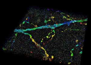
This image shows a 3D super-resolution reconstruction of dendrites in primary visual cortex.
Since Robert Hooke’s first description of a cell in Micrographia 350 years ago, microscopy has played an important role in understanding the rules of life.
However, the smallest resolvable feature, the resolution, is restricted by the wave nature of light. This century-old barrier has restricted understanding of cellular functions, interactions and dynamics, particularly at the sub-micron to nanometer scale.
Super-resolution fluorescence microscopy overcomes this fundamental limit, offering up to tenfold improvement in resolution, and allows scientists to visualize the inner workings of cells and biomolecules at unprecedented spatial resolution.
Such resolving capability is impeded, however, when observing inside whole-cell or tissue specimens, such as the ones often analyzed during the studies of the cancer or the brain. Light signals, emitted from molecules inside a specimen, travel through different parts of cell or tissue structures at different speeds and result in aberrations, which will deteriorate the image.
Now, Purdue University researchers have developed a new technology to overcome this challenge.