 A team of researchers at Texas A&M University is developing a highly printable bioink as a platform to generate anatomical-scale functional tissues.
Dr. Akhilesh Gaharwar and the Texas A&M team are working together to develop these bioinks, known as nanoengineered ionic-covalent entanglement (NICE) bioinks that combine nonreinforcement and ionic-covalent network to provide more effective reinforcement, resulting in stronger structures, according to a news release. Results from a study were published in the American Chemical Society’s Applied Materials and Interfaces.
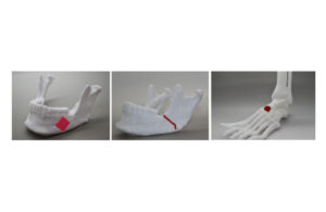
Once the bioprinting phase is complete, the cell-laden NICE networks are crosslinked, forming stronger scaffolds and allowing labs to produce full-scale, cell-friendly reconstructions of human body parts, including ears, blood vessels, cartilage and bone segments. The enclosed cells then deposit new proteins that calcify to form a mineralized bone over three months.
“The next milestone in 3D bioprinting is the maturation of bioprinted constructs toward the generation of functional tissues,” Gaharwar said in the release. “Our study demonstrates that NICE bioink developed in our lab can be used to engineer 3D-functional bone tissues.”
The researchers believe the technology could be applied in patient-specific bone grafts, developing replacement bone tissues to treat patients with arthritis, bone fractures, dental infections and craniofacial defects.
According to the release, Gaharwar and the team plan to demonstrate in-vivo functionality of the 3D-bioprinted bone tissue at some point in the future.