[Image from Duke University]
Duke University engineers are developing a fluid dynamics simulator that models blood flow in the full human arterial system at a subcellular resolution.
The biomedical engineers hope the simulator will give doctors guidance in treatment plans by simulating specific vasculatures of patients to predict how stent placements, conduit insertions and other geometric alterations to blood flow can affect surgical outcomes.
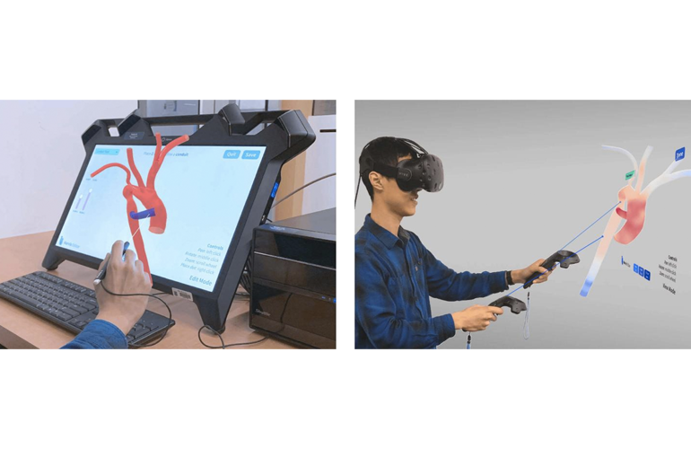
Duke University researchers developed the tool, called Harvey, by exploring various interfaces ranging from standard desktop displays to immersive virtual reality experiences. They found that futuristic interfaces could be the key to widespread adoption of the technology.
“Harvey currently requires knowledge of C coding and command-line interfaces, which really limits who can use the program,” Amanda Randles, assistant professor of biomedical sciences at Duke, said in a news release. “This paper introduces a graphical user interface we’ve developed called Harvey, so that anybody can use Harvey, whether they’re surgeons trying to figure out the best placement for a stent or biomedical researchers trying to design a new type of stent altogether.”
Randles said she has been developing the Harvey code for nearly 10 years and has demonstrated that the technology can accurately model blood flow through patient-specific aortas and other vascular geometries on longer scales. The program has also modeled 3D blood flows on the scale of the full human body.
Through the research, the simulation is bringing a better understanding of stent treatment of cerebral aneurysms and the growth of aneurysms. Randles also created a quick noninvasive way to check for peripheral arterial disease to understand how circulating cancer cells stick to different tissues.
“As cardiovascular disease continues to be the number one cause of death in the US, the ability to improve treatment planning and outcome remains a significant challenge,” said Randles. “With the maturity and availability of VR/AR devices, we need to understand the role these technologies can play in the interaction with such data. This research is a much-needed step for developing future software to combat cardiovascular disease.”
In a new study, Randles and her team of biomedical engineers are simulating three different scenarios to test the system: placing a conduit between two blood vessels, expanding or shrinking the size of a blood vessel and placing a stent within a blood vessel. The researchers attempted each simulation using a standard mouse and computer screen, a “Z-space” semi-immersive virtual reality device and a fully immersive virtual reality experience with an HTC Vive display device.
According to the researchers, the study didn’t find any major differences between the most and least immersive interfaces in terms of quality and efficiency.
“People enjoyed the 3D interface more,” said Randles. “And if they enjoyed it more, they’re more likely to actually use it. It could also be a fun and exciting way to get students engaged in classes about the vasculature system and hemodynamics.”
The researchers plan to run experiments to see if the 3D blood flow interface can help medical students retain important knowledge better than current standards.
The research was published in the Journal of Computational Science.