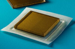
MIT engineers have developed a postage-stamp-sized, wearable ultrasound device. [Photo courtesy of MIT]
A team of MIT engineers is working to make getting an ultrasound as simple as buying a book of stamps.
They’ve developed a postage-stamp-sized ultrasound device that can be stuck to the skin and worn for continuous imaging of internal organs over 48 hours.
Once the engineers work out wireless connectivity, the ultrasound stickers could be worn by patients at home or on the go, even during exercises like jogging, biking and lifting weights, they said as they presented their work in Science. The ultrasound stickers could be used to monitor internal organs, tumor progression, fetal development or even the point of a workout regimen where further exertion will lead to muscle overuse and soreness.
“We envision a few patches adhered to different locations on the body, and the patches would communicate with your cellphone, where AI algorithms would analyze the images on demand,” Xuanhe Zhao, a professor of mechanical engineering and civil and environmental engineering at MIT, said in a news release. “We believe we’ve opened a new era of wearable imaging: With a few patches on your body, you could see your internal organs.”
Zhao was the study’s senior author, joined by lead authors Chonghe Wang and Xiaoyu Chen, and co-authors Liu Wang, Mitsutoshi Makihata, and Tao Zhao at MIT, plus Mayo Clinic’s Hsiao-Chuan.
A wearable ultrasound imaging tool “would have huge potential in the future of clinical diagnosis,” Wang said in the release. “However, the resolution and imaging duration of existing ultrasound patches is relatively low, and they cannot image deep organs.”
The MIT ultrasound sticker is about 3 mm thick and has a stretchy adhesive layer with a rigid array of transducers, which allows the device to conform to the skin so it can generate clearer, more precise images, Wang said. Two layers of elastomer in the adhesive layer encapsulate a middle layer of solid, water-based hydrogel for sound wave transmission.
“The elastomer prevents dehydration of hydrogel,” Chen said. “Only when hydrogel is highly hydrated can acoustic waves penetrate effectively and give high-resolution imaging of internal organs.”
The researchers used their prototypes on healthy individuals and watched their blood vessels change diameter when seated and standing, saw the stomach stretching and shrinking as volunteers drank and passed juice, observed the heart changing shape during exercise and even spotted bright patterns indicating microdamage in muscles during workouts.
In addition to the wireless efforts underway, the team is developing software algorithms to interpret the ultrasound imagery and offer diagnoses.
“We imagine we could have a box of stickers, each designed to image a different location of the body,” Zhao said. “We believe this represents a breakthrough in wearable devices and medical imaging.”