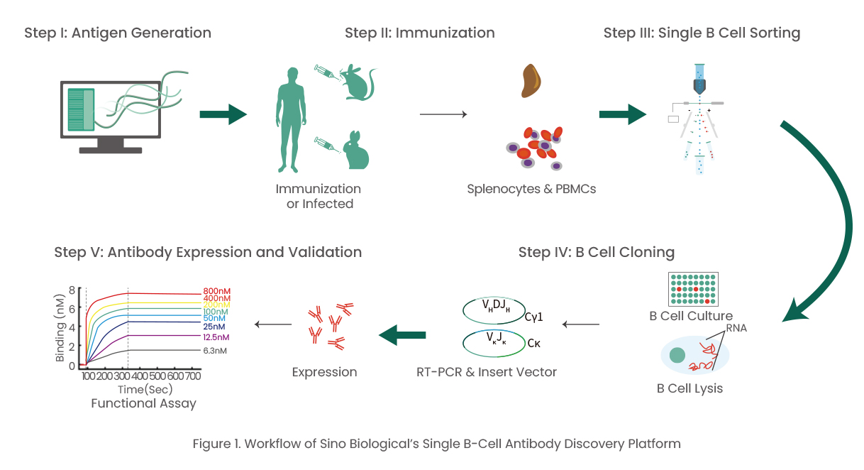
Introduction
Since the approval of Orthoclone OKT3 in 1986, more than 100 monoclonal antibodies (mAbs) have been approved by the Food and Drug Administration (FDA) to treat a variety of diseases ranging from autoimmune disorders, infectious diseases, and cancer [1-2]. In the context of the current COVID-19 pandemic, it’s crucial that these biologics are developed rapidly and efficiently. Among various antibody discovery approaches, including hybridoma technology, single B cell screening is a powerful and efficient strategy for generating antigen-specific mAbs based on the direct amplification of the VH and VL regions encoding genes from single B cells [3-4]. Notably, single B cell screening has various advantages that include maintaining the naïve VH/VL pairing, requiring relatively few cells, and the ability to discover antibodies against challenging targets.
Single B Cell Screening Technologies
Single B cell screening technologies have emerged and evolved rapidly over a decade. The process involves single B cell isolation, culture, sequencing, and cloning and the screening of selected single B cell antibodies. At the initial B cell screening stage, multiple platforms, including Berkely Lights Beacon®, fluorescence- activated cell sorting (FACS), and bead-based or FRET-based technologies, are applied. At Sino Biological, our team has established mature Beacon platform and flow cytometry platform for high throughput screening.
Single B Cell Screening using FACS
FACS is usually used by researchers to isolate single cells because of its high throughput capacity and sensitivity. The principle underlying B cell sorting is the clear distinction between antigen-specific B cells and other B cell subsets based on specific cell surface markers [5]. B cells are screened and sorted using flow cytometry after they are harvested from immunized animals or patients. If the flow cytometry panel is available, cells can be distinguished and sorted at different stages of development and differentiation based on the specific cell surface marker expression. Class-switch memory B cells and antibody-secreting cells (ASCs) are of interest to the researchers. Using fluorescence-labeled antibodies targeting CD19, IgM, IgD, IgG1, and antigens, B cells that exhibit high binding affinity can be selected. The selected cells are subjected to lysis directly after sorting in preparation for sequencing or they can be cultured and further screened using an ELISA with the culture supernatant prior to cell lysis.
Single B Cell Screening using Berkely Lights Beacon®
Using Beacon technology involves placing thousands of activated memory B cells and ASCs into NanoPen® chambers. A supernatant containing secreted antibodies reacts with the antigen-coated beads, surrounded by fluorescence-conjugated secondary antibodies. Fluorescence then indicates the antigen-specific antibodies and desired clones. Screening several thousands of clones takes minutes to hours. The selected clones are exported and retrieved for lysis using a structured light-based system called OptoSelect™ OEP (opto-electric positioning).
Advantages and Disadvantages
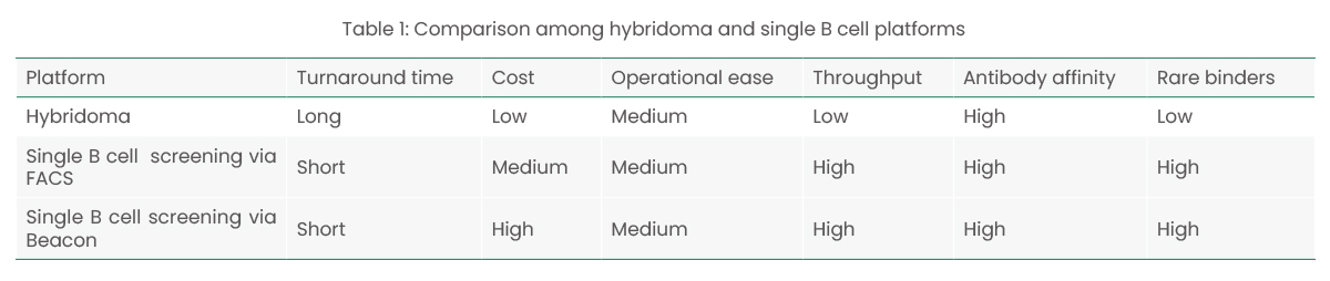
Compared with traditional hybridoma methods, single B cell screening platforms deliver different research and development (R&D) and production modes. Both traditional and single B cell methods provide naïve VH/VL pairing, but single B cell platforms are able to screen several thousand individual clones within a short period, making it possible to isolate rare binders. The R&D costs are higher with single B screening technology because of the requirement. (Table 1)
Case Study: Checkpoint Inhibitors Discovery for TIM-3
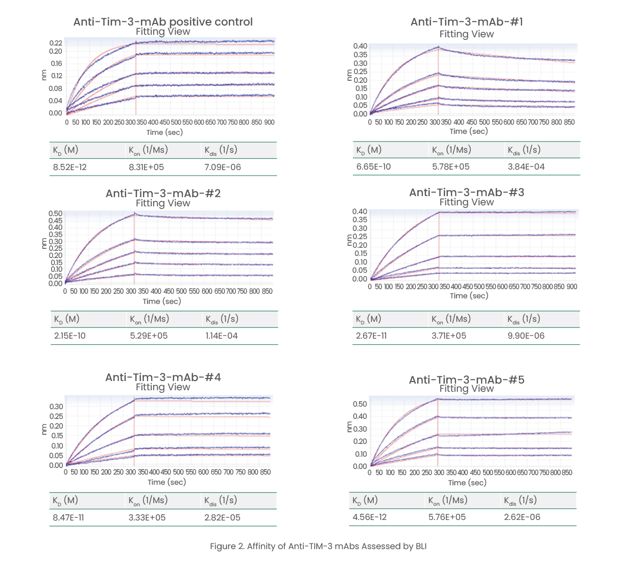
Using single B cell screening platforms, Sino Biological has developed anti-TIM-3 monoclonal antibodies with high affinity and neutralizing activity. After cell sorting and proliferation, 200 antigen-specific antibodies were identified from 1,100 single B cell supernatants. The kinetics and affinity of 63 purified ELISA-positive anti-TIM-3 monoclonal antibodies were evaluated using biolayer interferometry (BLI) technology. Among the ELISA-positive antibodies evaluated, five showed high affinity between 1010 and 1012 M (Figure 2). Five anti-TIM-3 mAbs were found to block the interaction between TIM-3 and PtdSer efficiently and in a dose-dependent manner, with an IC50 of 0.070 ± 0.010 μg/mL that was close to that of the positive control (Figure 3).

References
[1] DOI: 10.1016/j.intimp.2021.108112
[2] DOI: 10.1186/s12929-019-0592-z
[3] DOI: 10.1007/978-1-4939-8958-4_5
[4] DOI: 10.1016/j.nbt.2011.03.014
[5] DOI: 10.1371/journal.pone.0152282
Sponsored content by Sino Biological